History of Non-invasive Pacing
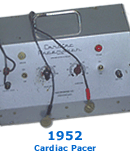
The concept of non-invasive cardiac pacing has been known for about 200 years. In 1791, Galvani reported that an electrical current applied across the heart of a dead frog resulted in myocardial contraction. However, non-invasive pacing was not made practical until Dr. Paul Zoll's work in the early 1950s. In 1952, Dr. Zoll reported the successful delivery of current from a generator through subcutaneous needle electrodes, which resulted in fixed rate pacing of two patients with ventricular standstill.1 He later reported the development and successful use of the first true non-invasive pacemaker and monitor. This device used a pair of 3-centimeter diameter metal electrodes secured to the chest wall and delivered 2-millisecond, 120-volt, alternating current impulses. Dr. Zoll is the acknowledged father of external pacing due to these early achievements.

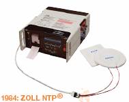
Technological improvements during the early 1980s made non-invasive pacing more comfortable and less cumbersome than earlier efforts. In 1981, ZOLL Medical Corporation patented and introduced a non-invasive external pacemaker with a longer pulse duration (40 milliseconds) and a larger electrode surface area (80 cm2). With stimuli of 40-millisecond duration, the threshold for cardiac responses was reduced, usually to 35-70 milliamperes (mA). A larger electrode surface area decreased the current density. Both of these factors greatly reduced the extent and severity of muscle contractions, and thus decreased the related discomfort. With the development of high impedance electrode gel on disposable electrodes, the current density was further decreased across the skin, eliminating cutaneous nerve pain.
In this new generation NTP, the electrocardiogram (ECG) monitor presented a clearly recognizable symbol of the stimulus artifact, which precisely marked the time of stimulation. It was designed with a “blanking period” of 80 milliseconds right after the stimulus signal so that the ECG was not impacted and ECG complexes were recognizable during pacing. Another advantage of the new external pacing device was its ability to operate in the demand mode (VVI), whereas older units were fixed rate (VOO).2

In 1982, the FDA approved the ZOLL non-invasive pacing device for use in patients with heart rates less than 40 beats per minute and asystole. Other manufacturers have subsequently developed similar devices, now most often combined with defibrillators.
Physiology of Non-invasive Transcutaneous Pacing
When a strong electrical current is applied to the chest with NTP, a small amount will reach the heart, stimulating it to depolarize and contract. In transcutaneous pacing, the right and left ventricles are stimulated. Frequently the atria are activated by retrograde conduction. There is atrioventricular dissociation, so the atrial kick is lost, resulting in an approximately 20% decrease in cardiac output. For a patient with symptomatic bradycardia, NTP results in an increased cardiac output, increased mean arterial pressure, and decreased systemic vascular resistance. Studies have shown that with NTP, there is no elevation in Creatine Kinase, in muscle and brain tissue, so there is no myocardial damage. Additionally, it was found that the hemodynamic response was similar for NTP and transvenous ventricular endocardial pacing.
When ECG electrodes are attached for sensing of the QRS, the pacemaker is able to operate in the demand
mode. A pacemaker stimulus will occur if the intrinsic signal is slower than the pacing rate programmed by the clinician. If the device senses that the patient’s heart rate is faster than the pacing rate, it inhibits the pacemaker’s electrical signal.
Asynchronous (fixed rate) pacing, can also be selected on the noninvasive device. In this mode the device paces at the rate set by the clinician, independent of the patient’s intrinsic heart rhythm. Asynchronous pacing is used when:
- There is not time to put ECG electrodes in place.
- When the pacemaker is inhibited due to sensing of signals other than the R wave, e.g., artifact from patient movement, P or T waves, or interference from another electrical device.
During asynchronous pacing, there may be competition between the patient’s own beats and the paced beats, unless asystole is present. There is the potential for the pacemaker stimulus to land on the T wave during the vulnerable period. Refer to the section Safety of Noninvasive Transcutaneous Pacing for the significance of this factor.
1. Zoll, PM. Resuscitation of heart in ventricular standstill by external electrical stimulation. NEJM. 1952;247:768-771.
2. Worley SJ, Bride WM. External transthoracic pacing in patients with acute myocardial infarction. In: RM Califf, GS Wagner, (Eds) Acute Coronary Care. Boston: Martinus Nijhoff; 1987.